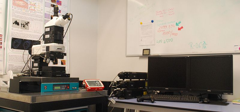
We perform Raman measurements of bio-samples (incl. cells and tissues), materials, and chemicals, employing Confocal WITec Alpha300 R spectrometer coupled with atomic force microscope AFM. Samples can be examined in air, solid state, and in solutions in a non-invasive manner.
The system is equipped with:
- 3 lasers: 488, 532, and 633 nm;
- air objectives: 20×, 40×, 100×;
- immersive objectives: 20×, 40×, 63×;
- EMCCD detector (time of measurement for a single spectrum of 1 ms);
- power control of the excitation radiation (with an accuracy of 0.1 µW);
- automatic table (range of motion: 20 mm x 20 mm (X-Y planes), positioning of sample with precision up to 10 nm).
- Confocal Raman measurements can be performed in 3 modes: single measurements, along the line in the X-Y and Z planes (2D imaging), and 3D imaging with spatial resolution limited by the diffraction limit (lateral > 300 nm, axial > 700 nm) and spectral resolution up to 0.5 cm-1.
Thanks to the AFM microscope the imaging of samples in contact (for hard samples) and intermittent contact (for soft samples) modes is possible. We perform the measurements of topography and mechanical properties i.e. stiffness and adhesion. Additionally, the correlation of the Raman image with the AFM image (measurements without transferring the sample) can be executed.
We also perform Raman measurements employing Confocal WITec Alpha300 RS coupled with AFM and Scanning Near-Field Optical Microscope (SNOM). The design of this Alpha300 RS setup features a Confocal Microscope (CM), a Scanning Near-Field Optical Microscope (SNOM) and an Atomic Force Microscope (AFM) in a single instrument.
This RS setup is equipped with lasers: 405, 488, and 532 nm.
SNOM measurements can be performed in bottom up, top down, and collection modes. Optical imaging of sample surface can be realized with resolution beyond the diffraction limit. Additionally, the combination of high-resolution surface imaging (SNOM) and chemical imaging (Raman) is possible.
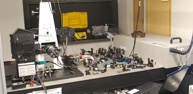
We perform Raman imaging employing a home-built SRS system, the first SRS system in Poland and a few in the world!
The central part of the system is the Fluence Lazurite pulsed 2 ps laser with 20 MHz's of repetition rate that is based on fiber laser producing 1029 nm Stokes beam with 450 mW of average power with the built-in optical parametric oscillator (OPO) that produces tunable wavelength beam in the range of 750–950 nm with an average power of 100 mW. SRS microscope enables measurements in the spectral range of 3600-1000 cm-1, i.e. both in the range of C-H vibrations (identification of lipids, proteins – 3600-2700 cm-1) and in the so-called “fingerprint region” (<1800 cm-1). The "silent" range (2700-1800 cm-1) is very helpful in tracking various exogenous substances that have characteristic structures that give a Raman signal in this regard.
The system is currently being extended for spontaneous Raman (RS) and Coherent anti-Stokes Raman (CARS) microscopy.

We perform FT-IR measurements of bio-samples (incl. cells, tissues, and plants), materials, and chemicals, employing Agilent 670-IR spectrometer combined with the 620-IR microscope 620-IR. Samples can be examined in air, solid state, and in solutions in a non-invasive manner.
The system is equipped with:
- Cassegrain IR objectives: magnification of 15× and 25×;
- single element MCT detector (spectral region of 670-4000 cm-1);
- Focal Plane Array (FPA) detector cooled with liquid nitrogen (spectral region of 850-4000 cm-1) – consists of a matrix of 128×128 pixels giving 16 384 spectra simultaneously;
- Agilent optical setup for 5-fold enhancement of spatial resolution;
- automatic table.
- FT-IR measurements can be performed in 3 modes: transmission, reflection and ATR (depending on sample properties). Spectra acquisition is realized through single measurements or FT-IR imaging (mosaic mode) with following definitions:
- Standard definition imaging (pixel size: 5.5×5.5 µm2) from an area of 700×700 µm2;
- HD – high definition imaging (pixel size: 1.1×1.1 µm2) from an area of 140×140 µm2;
- UHD – ultra-high definition imaging (pixel size: 0.66×0.66 µm2) from an area of 85×85 µm2;
- ATR imaging (pixel size: 1.1×1.1 µm2) from an area of 70×70 µm2.
The system has built-in ATR-FT-IR attachment for collection of single FT-IR spectra. ATR-FT-IR setup is equipped with single reflection diamond crystal (spectral region of 375-4000 cm-1 and spectral resolution from 0,5 to 16 cm-1).
Additionally, we also own an original CorrescopyTM system, which enables the registration of the RS and FTIR maps from identical areas of sample. Therefore, the direct comparison of collected maps is possible.
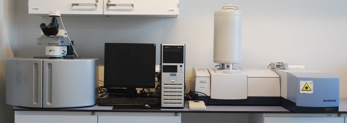
We perform Raman measurement of bio-samples, materials, pharmaceuticals, powders, and chemicals employing MultiRAM FT-Raman Bruker spectrometer coupled with RamanScopeIII microscope. Samples can be examined in air, solid state, and in solutions in a non-invasive manner.
The system is equipped with:
- laser: 1064 nm;
- air objectives: 10×, 40× (with increment of ca. 1 µm);
- Ge detector cooled with liquid nitrogen;
- temperature-controlled device;
- automatic table for mapping in XYZ planes (with an increment of ca. 5 μm).
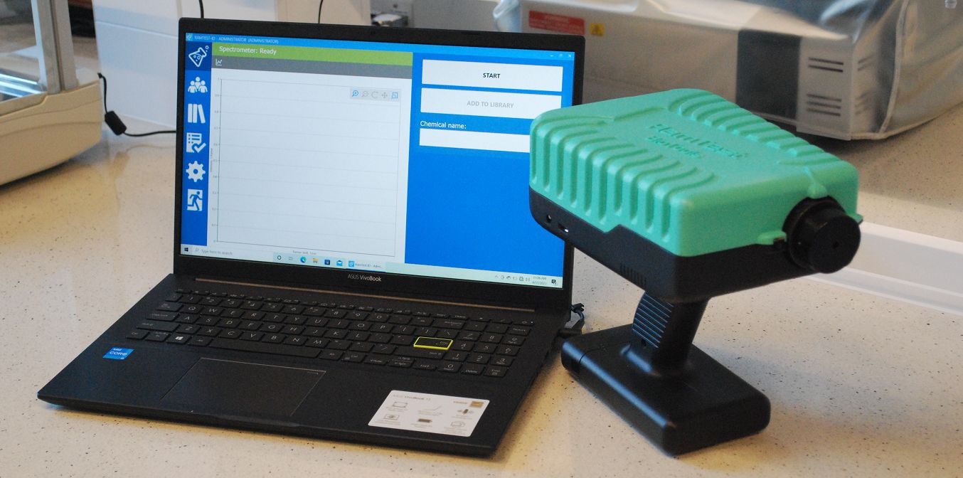
We perform Raman measurements of bio-samples and chemicals employing portable RamTestTM spectrometer. The RamTestTM setup is a modern handheld instrument that enables in-situ sample inspection.
The setup is equipped with a 532 nm laser excitation (unique for portable RS). The measurements can be realized in a spectral range of 100-4000 cm-1 in a rapid manner. The setup has built in automatic fluorescence and baseline correction which enables the acquisition of high-quality spectra.
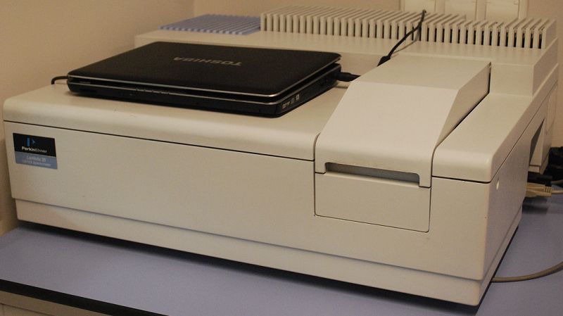
We perform UV-VIS-NIR measurements with Perkin Elmer Lambda 25 spectrophotometer equipped with:
- deuterium and Tungsten realigned sources with automatic switch-over (spectral range: 190 - 1100 nm),
- double beam,
- sealed, quartz-coated mirrors,
- lens-free system to reduce chromatic aberrations,
- monochromator (Seya Namioka).
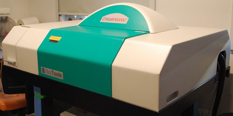
ROA is a form of Vibrational Optical Activity (VOA) that is complementary to Vibrational Circular Dichroism (VCD) in the same way that Raman spectroscopy is complementary to IR spectroscopy. ROA is defined as the difference in Raman intensity for left (L)- minus right (R)-circularly polarized incident and/or scattered radiation in chiral molecules. It combines the structural specificity of vibrational spectroscopy with the stero-sensitivity of chiral detection. We perform ROA measurement employing ChiralRAMAN BioTools Inc. and ChiralRAMAN-2x BioTools Inc. ROA spectrometers.
The setups are equipped with:
- 2 W green diode laser 532 nm, with adjustable power output;
- CCDsp camera Critical Link detector;
- SCP configuration (Scattered Circular Polarization);
The measurements of Raman/ROA spectra can be realized with ChiralRAMAN and ChiralRAMAN-2x in spectral regions of 200-2500 cm-1 and 100-2500 cm-1, respectively. The latter is additionally equipped with temperature control, in range of -5°C-80°C.
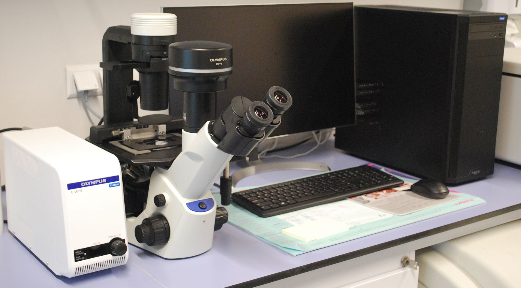
We collect fluorescence images of bio-samples employing optical microscope Olympus CKX53.
The setup is equipped with:
- a high-brightness U-LGPS light source to excite fluorescence;
- set of excitation filters adjusted to selected dyes;
- air objectives: 10×, 20×, 40×;
- high-resolution camera (Olympus DP74);
- CellSens software.
The images obtained can be saved in TIFF format.
Our newly established laboratory is equipped with all necessary equipment for biological experiments :
- Alpina Bio130 laminar chamber with the possibility of connecting to a vacuum, additionally equipped with a Biovac suction system;
- 2 incubators: 180 L (Safegrow Pro) and 50 L (ESCO);
- -80˚ C freezer (Vestrfrost);
- a dewar (Auguste Cryogenics) for cell culture banks storage;
- a standard fridge-freezer;
- an autoclave (Tuttnauer);
- hemocytometer;
- 2 centrifuges (MPW).