Przejdź do głównej treści

Widok zawartości stron Widok zawartości stron

Pomiń baner

Widok zawartości stron Widok zawartości stron

Widok zawartości stron Widok zawartości stron

RSC Molecular Spectroscopy Group Online Seminar Series - October 2021 (21.10.2021)

On 21st October 2021 dr Anna Nowakowska, gave a presentation entitled Towards clinical diagnostics - can Raman spectroscopy detect and distinguish leukemia cells? during the RSC Molecular Spectroscopy Group Online Seminar Series organized by the Royal Society of Chemistry.

Form: oral presentation

więcej o

Widok zawartości stron Widok zawartości stron

17th Confocal Raman Imaging Symposium (27.09-01.10.2021)- Abstracts

Accumulation of lipids as a marker of T cell activation revealed by label-free Raman spectroscopy

Aleksandra Borek-Dorosz1,2, Anna Maria Nowakowska1, Paulina Laskowska3, Maciej Szydłowski3, Małgorzata Zasowska3, Przemysław Juszczyński3, Maciej Szydłowki3, Piotr Mrówka3,4, Malgorzata Baranska1,2, Katarzyna Majzner1,2*

1Jagiellonian University, Faculty of Chemistry, Krakow, Poland

2Jagiellonian University, Jagiellonian Centre for Experimental Therapeutics (JCET), Krakow, Poland

3Department of Experimental  Hematology, Institute of Hematology and Transfusion Medicine, Warsaw, Poland

4Department of Biophysics, Physiology and Pathophysiology, Medical University of Warsaw, Warsaw, Poland

T cells are one of the most important white blood cells of the immune system and play a central role in the adaptive immune response. T cells are originated from pluripotential hematopoietic stem cells [1]. In order to become fully functional effector cells, the activation requires specific antigens, binding to receptors presented in T-cell (TCR). Another requirement is the co-stimulating interaction of other surface proteins and cytokines. Activating signal leads to a cascade of intracellular events resulting in a production of subtype-specific effector proteins and acceleration of proliferation [2]. A deeper understanding of biochemical changes followed by T cells activation will allow the improvement of immunotherapy and CAR-T treatment. Therefore, the goal of the studies was the identification of molecular changes triggered by the activation of T cells by means of Raman imaging.

In our studies, we applied label-free Raman imaging for molecular characterization and discrimination of naive and activated T cells. We have defined spectral biomarkers characterizing the activation process, such as increased concentration of lipids in the cell cytoplasm, changes in the nucleus morphology and chromatin condensation and decrease of carotenoids accumulation. These molecular changes were clearly evidenced in the Raman spectra collected from naïve and activated T lymphocytes and proved by multivariate analysis, which was carried out (Fig. 1). The reliable recognition of activated T cells based on the Raman spectra was possible after a detailed analysis of the average spectra and application of principal components analysis (PCA) and partial least squares discrimination analysis (PLS-DA) methods.

Acknowledgments

This work was supported by „Label-free and rapid optical imaging, detection and sorting of leukemia cells” project carried out within the Team-Net program of the Foundation for Polish Science co-financed by the EU under the ERDF.

References
[1] 5. Hematopoietic Stem Cells | stemcells.nih.gov". stemcells.nih.gov. Retrieved 2020-11-21.
[2] D. G. Waller, A. P. Sampson (2018) Rheumatoid arthritis, other inflammatory arthritides and osteoarthritis,  Medical Pharmacology and Therapeutics (Fifth Edition), Elsevier

Form: poster presentation

więcej o

Molecular Raman Probes-based detection of promieloblastic cells  biochemical state

Adriana Adamczyk1, Anna M. Nowakowska1, Katarzyna Majzner1,2, Małgorzata Barańska1,2

1Jagiellonian University, Faculty of Chemistry, Krakow, Poland

2Jagiellonian University, Jagiellonian Centre for Experimental Therapeutics (JCET), Krakow, Poland

Raman Spectroscopy is recognized as a label-free method that brings multiple advantages for the characterization of biological samples. Biochemical processes, also linked to pathology development, are associated with dysfunctions of cellular organelles. Marker bands of biocomponents such as DNA, phospholipids, and cytochrome C characterize the biochemical state of crucial cellular compartments: nucleus, endoplasmic reticulum, and mitochondria, respectively. However, the lack of marker bands of some cellular structures or difficulties in the detection outlines the need to develop molecular Raman probes. The combination of selective targeting moiety and a reporting part containing triple bonds or deuterium substitution and displaying signal in 1800-2800 cm-1 range enable simultaneous detection of multiple probes without bands overlapping with a compound of cellular origin. Application of Raman probes for recently developing non-linear Raman-based techniques, benefiting in signal increase, could provide an innovative alternative to existing diagnostic methods.1

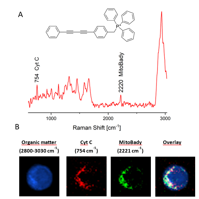
The ultimate goal of the study was to evaluate drug-induced granulocytic differentiation of promyelocytic cells towards neutrophile-like cells using the HL-60 cell line. Changes associated with mitochondria state were followed by Raman imaging using MitoBADY probe (Fig.1), displaying band at 2220 cm-1‑ that accumulates in negatively charged mitochondria membrane, thanks to the positive charge of the compound. We proved that MitoBADY is mitochondria specific probe. Even low concentrations of MitoBADY (100 nM) and low incubation time (15 min) are enough to characterize mitochondria distribution in cells.

The „Label-free and rapid optical imaging, detection and sorting of leukemia cells” project is carried out within the Team-Net programme of the Foundation for Polish Science co-financed by the EU.

1.           Adamczyk, A. et al. Toward Raman Subcellular Imaging of Endothelial Dysfunction. J. Med. Chem. (2021) doi:10.1021/acs.jmedchem.1c00051.

Form: poster presentation

więcej o

Biochemical characterization and discrimination of B-type acute lymphoblastic leukemia (B-ALL) by Raman imaging

Patrycja Leszczenko1, Aleksandra Borek-Dorosz1,2, Anna Maria Nowakowska1, Adriana Adamczyk1, Sviatlana Kashyrskaya1, Justyna Jakubowska3, Marta Ząbczyńska3, Agata Pastorczak3, Kinga Ostrowska3, Małgorzata Barańska1,2, Katarzyna Maria Marzec2, Katarzyna Majzner1,2

1Faculty of Chemistry, Jagiellonian University, Krakow, Poland;

2Jagiellonian Centre for Experimental Therapeutics (JCET), Jagiellonian University, Krakow, Poland;

3Medical University of Łódź, Department of Pediatrics, Oncology and Hematology, Łódź, Poland

Acute lymphoblastic leukemia (ALL) is the most common childhood malignancy. B-type ALL (B-ALL) originate from immature B cell precursors. The ALL subtype, characterized by the detected fusion gene, influences the prognosis and the effectiveness of the therapy [1]. Currently used diagnostic methods are expensive and time-consuming. Therefore, seeking for novel diagnostic methods of blood malignancies is still unflagging need within oncology prospect. Raman spectroscopy (RS) is a rapid technique providing spatial resolution at the subcellular level, thereby supplying information about the biochemical composition of the sample. RS due to many advantages might be considered as a potential and valuable diagnostic method over the next years [2, 3].

In our studies, we used Raman imaging supported with chemometrics analysis, in order to effectively distinguish healthy B cells from their leukemic counterparts. Samples of three subtypes of B-ALL (BCR-ABL1, TCF3-PBX1, TEL-AML1), were isolated from the bone marrow of diagnosed patients, whereas B cells were derived from whole peripheral blood of healthy donors. Samples were measured by RS with excitation at 532 nm and 633 nm, resulting in a total number of spectra above 153 600. Raman images of single cells were analysed using k-means cluster analysis (KMCA). Principal component analysis (PCA) allowed us to reveal the subtle differences between the spectra of studied cells. We discovered that the Raman spectra of healthy B cells were heavily influenced by signals attributed to nucleic acids and proteins. Partial least squares (PLS) regression was used to establish an algorithm to differentiate spectra of healthy B lymphocytes from leukemic cells. The method delivered prosperous results with high accuracy. However, designing the algorithm to distinguish spectra of all studied subtypes of B-ALL was not possible. Despite difficulties, we managed to obtain discrimination between BCR-ABL1 and TCF3-PBX1 samples, proving that spectral determination of molecular subtypes of B-ALL is possible. Presented results demonstrate the potential of RS in combination with chemometric analysis in the diagnosis of leukemia in clinical practice.

[1] Larson, R.A. Acute lymphoblastic leukemia. In Williams Hematology; McGraw-Hill Education, 2016; pp. 1505–1526 ISBN 978-0-07-183301-1

[2] Managò, S.; Valente, C.; Mirabelli, P.; De Luca, A.C. Discrimination and classification of acute lymphoblastic leukemia cells by Raman spectroscopy. Opt. Sensors 2015 2015, 9506, 95060Z, doi:10.1117/12.2179486

[3] Hassoun, M.; Köse, N.; Kiselev, R.; Kirchberger-Tolstik, T.; Schie, I.W.; Krafft, C.; Popp, J. Quantitation of acute monocytic leukemia cells spiked in control monocytes using surface-enhanced Raman spectroscopy. Anal. Methods 2018, 10, 2785–2791

Acknowledges

The „Label-free and rapid optical imaging, detection and sorting of leukemia cells” project is carried out within the Team-Net programme of the Foundation for Polish Science co-financed by the EU.

Form: poster presentation

więcej o

Spectroscopic characterization of IDH1 and IDH2- mutated transgenic HEL cells

Anna M. Nowakowska1,  Paulina Laskowska2, Aleksandra Borek-Dorosz1,3, Adriana Adamczyk1, Patrycja Leszczenko1, Małgorzata Zasowska2, Maciej Szydłowski2, Piotr Mrówka2,4, Małgorzata Barańska1,3, Katarzyna Majzner1,3

1Faculty of Chemistry, Jagiellonian University, Krakow, Poland

2Department of Experimental  Hematology, Institute of Hematology and Transfusion Medicine, Warsaw, Poland

3Jagiellonian Centre for Experimental Therapeutics (JCET), Jagiellonian University, Krakow, Poland

4Department of Biophysics, Physiology and Pathophysiology, Medical University of Warsaw, Warsaw, Poland

Abnormalities occurring in the genome of the precursors of different blood cells lead to the development of leukemia. Different subtypes of cancer of the blood cells are defined by specific gene alterations influencing phenotype, aggressiveness, and drug resistance of cancer cells. Nowadays, new in vitro models are now being sought to enable testing of new anti-cancer drugs and deepening molecular characterization of leukemic cells. Some of the genes that are frequently mutated in leukemias are IDH1 and IDH2 genes, encoding isocitrate dehydrogenase 1 and 2 [1]. Therefore biochemical characterization of transgenic cell lines with IDH1 and IDH2 gene rearrangements is desired in the context of studies of leukemia development.

The goal of our study was the identification of metabolic changes associated with IDH1 and IDH2 mutations in HEL cells based in vitro model of leukemia using Raman imaging combined with multivariate statistical analysis [2]. We focused on studies of two mutant HEL cell lines related to rearrangements of IDH1 and IDH2 genes, resulting in a replacement of arginine with histidine at the residue 132 (IDH1/R132H) or in a replacement of arginine with glutamine at the residue 140 (IDH2/R140Q). As a result of gene alterations of IDH1 and IDH2 genes, loss of their normal catalytic activity is observed. Indirectly, it leads to abnormal histone and DNA methylation and causes alterations in the differentiation of progenitor cells and neoplasm development. HEL cells with a wild-type (WT) sequence of IDH1 and IDH2 genes served as control samples. Control and IDH-mutated transgenic cells were imaged using the confocal Raman system WITec Alpha 300. Obtained Raman maps were subjected to k-means cluster analysis (KMCA). Obtained mean spectra of cells and cellular components were further analyzed using principal components analysis (PCA) and partial least squares regression (PLS) chemometric methods in order to identify subtle differences of HEL cells carrying different variants of IDH1 and IDH2 genes. Mutated and parental HEL cells primarily differed in the protein and nucleic acids composition.

Acknowledges:

The „Label-free and rapid optical imaging, detection and sorting of leukemia cells” project is carried out within the Team-Net programme of the Foundation for Polish Science co-financed by the EU.

[1] H. Yang, D. Ye, K-L. Guan, Y. Xiong, IDH1 and IDH2 mutations in tumorigenesis: mechanistic insights and clinical perspectives, Clin Cancer Res, 2012, DOI: 10.1158/1078-0432.CCR-12-1773.

[2] P. Martin and T. Papayannopoulou, HEL cells: a new human erythroleukemia cell line with spontaneous and induced globin expression, Science, 1982,
DOI: 10.1126/science.6177045

Form: poster presentation

więcej o

Widok zawartości stron Widok zawartości stron

29th Congress of the Polish Society of Hematologists and Transfusionists / XXIX Zjazd Polskiego Towarzystwa Hematologów i Transfuzjologów - (02-03.09.2021)

Paulina Laskowska from the Department of Experimental  Hematology, Institute of Hematology and Transfusion Medicine, Warsaw, Poland, who collaborate with us, gave a presentation entitled Identification of activated T cells using Raman-based methodology during the 29th Congress of the Polish Society of Hematologists and Transfusionists.

Form: oral presentation


więcej o

Widok zawartości stron Widok zawartości stron

11th International Conference on Advanced Vibrational Spectroscopy (ICAVS, 23-26.08.2021)- Abstracts

11th International Conference on Advanced Vibrational Spectroscopy (ICAVS, 23-26.08.2021)- Abstracts

Raman spectroscopic differentiation between human peripheral blood mononuclear cells and molecular characterization of naïve and activated T lymphocytes

Aleksandra Borek-Dorosz1,2, Anna Maria Nowakowska1, Patrycja Leszczenko1, Adriana Adamczyk1, Justyna Jakubowska3, Agata Pastorczak3, Kinga Ostrowska3, Marta Ząbczyńska3, Paulina Laskowska4, Maciej Szydłowski4, Małgorzata Zasowska4, Przemysław Juszczyński4, Piotr Mrówka4, Malgorzata Baranska1,2, Katarzyna Maria Marzec2*, Katarzyna Majzner1,2*

1Jagiellonian University, Faculty of Chemistry, Krakow, Poland

2Jagiellonian University, Jagiellonian Centre for Experimental Therapeutics (JCET), Krakow, Poland

3Medical University of Łódź, Department of Pediatrics, Oncology and Hematology, Łódź, Poland

4Department of Diagnostic Hematology, Institute of Hematology and Transfusion Medicine, Warsaw, Poland

Human peripheral blood mononuclear cells (PBMCs) are a heterogeneous population of cells that includes T, B lymphocytes, natural killer cells, monocytes, and dendritic cells. Lymphocytes represent the most numerous cell population within PBMCs (70-90%) [1] and are characterized by similar morphological features including size (~8-10 µm), large nucleus and cytoplasmic border containing subcellular structures [2].

Lymphocyte activation occurs when B or T cells are spark off through antigen-specific receptors on their cell surface that triggers proliferation and differentiation of cells into specialized effector lymphocytes. Activation of T lymphocytes involves multiple intracellular events, staring from recognition of specific antigen and co-stimulatory signal [3]. Activated T cells are important line of defence against pathogenic threat e.g. viral infection.

The total number of lymphocytes and their percentage within the blood can act as a marker for the diagnosis of diverse human diseases. Currently, methods based on cytometry are the most commonly used in order to distinguish subtypes of leukocyte and quantify their number. However, these techniques use cell immunophenotyping, which is limited by the number of fluorochrome-labeled antibodies that can be applied simultaneously. In our studies we applied a label-free Raman spectroscopy imaging method for molecular characterization and discrimination of PBMCs and activated T cells. We have defined spectral biomarkers characteristic for carotenoids, nucleic acids as well as proteins and lipids fractions, all detected and visualised on the sub-cellular level. We have presented that although the presence of carotenoids in lymphocytes T depends on the individual donor variability, the reliable distinction between PBMCs is possible based on the analysis of defined in this work Raman markers. The accumulation of carotenoids exclusively in lymphocytes T was clearly evidenced in the Raman spectra and supported by quantitative analysis carried out with the application of HPLC method. The reliable recognition was possible only after detailed analysis of the average spectra and application of PCA and PLS methods. 

Acknowledgments

This work was supported by „Label-free and rapid optical imaging, detection and sorting of leukemia cells” project carried out within the Team-Net program of the Foundation for Polish Science co-financed by the EU.

References

[1] Murphy K (2012) Janeway’s Immunobiology, 8th ed. Garland Science, Taylor & Francis Group, New York         
[2] Young NA, Al-Saleem T (2008) CHAPTER 24 - Lymph Nodes: Cytomorphology and Flow Cytometry. In: Bibbo M, Wilbur DBT-CC (Third E (eds). W.B. Saunders, Edinburgh, pp 671–711
[3] D. G. Waller, A. P. Sampson (2018) Rheumatoid arthritis, other inflammatory arthritides and osteoarthritis,  Medical Pharmacology and Therapeutics (Fifth Edition), Elsevier  

Form: flash presentation

więcej o

Raman reporters as a tool to evaluate promyeloblastic cells differentiation stage- optimization of imaging conditions.

Adriana Adamczyk1, Anna M. Nowakowska1, Katarzyna Majzner1,2, Małgorzata Barańska1,2

1Jagiellonian University, Faculty of Chemistry, Krakow, Poland

2Jagiellonian University, Jagiellonian Centre for Experimental Therapeutics (JCET), Krakow, Poland

Biochemical processes, also associated with pathology development, are recognized to be associated with cellular organelles dysfunctions.  For many years, studies on the single-organelle level were possible using fluorescence probes consisting of targeting moiety and reporting part that allows for signal detection. However, certain disadvantages, such as dyes photobleaching, cytotoxicity, or broad emission bands, triggered the development of organelle-specific probes for different spectroscopic techniques. Here, we focus on Raman spectroscopy that offers narrow spectral bands, sensitivity, and a possibility to maintain physiological conditions during measurements. Of particular interest is the spectroscopic region 1800-2800 cm-1, which displays no cell-specific vibrational bands, in opposition to deuterated or triple bond-containing compounds. Application of appropriately designed probes with intense bands in the cell silent region prevents possible overlapping of Raman signals and give better image contrast. The literature describes novel so-called Raman reporters based on triphenylphosphonium targeting moiety, called MitoBady (Fig. 1A) for mitochondria detection, thymine analogues 5-Ethynyl-2'-deoxyuridine (EdU) to study DNA replication and deuterated amino acids, fatty acids, etc.1 However, every external compound introduced to the cellular system might affect its function. In particular, compounds bearing charge, such as MitoBady, can affect cellular metabolism. Therefore, their cytotoxicity effect on cell homeostasis and appropriate Raman imaging conditions need to be assessed. The relevance of this approach could be additionally enhanced by the rapid development of non-linear Raman-based techniques, like stimulated Raman spectroscopy, that enables for a significant increase of signal intensity with simultaneously reduced integration time. High sensitivity, in combination with the specificity of Raman probes might  provide powerful diagnostic method for the early detection of multiple diseases.

Lymphocytes and myeloid lineages, originated from hematopoietic stem cells, are produced in bone marrow niches and developed towards well-functioning blood cells under specific conditions. In these studies, in vitro model of maturation towards neutrophils was investigated using human leukemia cell line Hl-60. Optimization of Raman imaging condition using MitoBady (Fig. 1B) and EdU allowed following changes associated with mitochondria and nucleus between undifferentiated promyeloblasts and mature neutrophils by detecting Raman signal of a living cell at 2120 and
2220 cm-1. The ultimate goal was to use Raman reporters to identify granulocytic differentiation pathologies during acute promyelocytic leukemia development.

Raman spectrum of HL-60 with MitoBADY

Figure 1 A- MitoBady structure and Raman spectrum of HL-60 cells mitochondria B- Distribution image of organic matter, cytochrome C, Mitbady, and images overlay.

The „Label-free and rapid optical imaging, detection and sorting of leukemia cells” project is carried out within the Team-Net program of the Foundation for Polish Science co-financed by the EU.


(1)      Adamczyk, A.; Matuszyk, E.; Radwan, B.; Rocchetti, S.; Chlopicki, S.; Baranska, M. Toward Raman Subcellular Imaging of Endothelial Dysfunction. J. Med. Chem. 2021. https://doi.org/10.1021/acs.jmedchem.1c00051.

Form: flash presentation

więcej o

Can Raman imaging classify abnormal B-lymphoblast of different origin? Spectroscopic studies of  MLL-rearranged, Philadelphia chromosome- positive and JAK2-mutated  B-cell precursor acute lymphoblastic leukemias

Adriana Adamczyk1, Aleksandra Dorosz1,2, Patrycja Leszczenko1, Sviatlana Kashyrskaya1, Agnieszka Grabacka1, Maja Bartoszek1, Anna M. Nowakowska1, Justyna Jakubowska3, Agata Pastorczak3, Kinga Ostrowska3, Marta Ząbczyńska3, Katarzyna Majzner1,2, Małgorzata Barańska1,2

1Jagiellonian University, Faculty of Chemistry, Krakow, Poland

2Jagiellonian University, Jagiellonian Centre for Experimental Therapeutics (JCET), Krakow, Poland

3Medical University of Łódź, Department of Pediatrics, Oncology and Hematology, Łódź, Poland

Acute lymphoblastic leukemia (ALL) results from impaired maturation and differentiation of lymphoid progenitors that invade bone marrow, peripheral blood and extramedullary sites . Several genetic alterations have been already found as drivers of ALL, uncovering the genetic heterogeneity of the disease. The presence of specific molecular aberrations including MLL gene rearrangements, BCR-ABL1 gene fusion (Philadelphia chromosome), and JAK2 mutations affect the response to conventional chemotherapy. .

Next to classical diagnostic techniques like immunophenotyping or karyotyping, Raman spectroscopy is considered as a new promising tool, which can support the diagnosis of leukemia in clinical practice.  Raman spectroscopy provides  many advantages in biological studies of cancer cells, including those resulting from  insignificant water bands intensity, high spatial resolution, photostability, sample non- destructiveness, high sensitivity and possibility to obtain full information of all the chemical components at once.1 Therefore, in our studies, we used confocal Raman imaging in combination with advanced chemometric analysis to classify pathological Blymphoblast representing different molecular subtypes of ALL. We performed Raman measurements of ALL cell lines with Philadelphia chromosome (Ph+, SUP-B15 cell line), harboring JAK2 point mutation (MHH-CALL4 cell line) and  MLL rearrangement (RS4;11 and SEM-K2 cell lines) with the use of two laser excitation wavelengths 532 nm and 633 nm in order to fully explore biochemical diversity of the cell lines studied. To obtain sufficient statistics, a minimum of 50 cells were measured from each sample and the experiment was repeated a minimum of three times. Our results show that in vitro models of three different subtypes of B-ALL leukemia representing B-lymphoblast with Ph+, MLL, or JAK2 gene mutations display characteristic spectroscopic features of biochemical cell state. Moreover, based on our studies, well discrimination can be observed between leukemia cells and normal blood lymphocytes, mainly based on the Raman signals characteristic for nucleic acids (eg. 790 cm-1 band). This overall picture demonstrates the diagnostic potential of Raman spectroscopy in combination with chemometrics analysis (k-means cluster analysis, principal component analysis, partial least squares regression) for the diagnosis of subtypes of leukemia in clinical practice. Moreover, detailed characterization of cellular metabolism is a key point for further Raman-based studies of already existing and new chemotherapeutic agents’ influence and effectiveness.

A scheme of the experiments and analysisFigure 1 Example of analysis scheme  of a single cell: Upper part: integration image at 3050-2800 cm-1 range, bottom part: a false colour image of whole-cell, cytoplasm and nucleus class and corresponding Raman spectra (laser line 532 nm)


The „Label-free and rapid optical imaging, detection and sorting of leukemia cells” project is carried out within the Team-Net program of the Foundation for Polish Science co-financed by the EU under the ERDF.

(1)         Managò, S.; Zito, G.; De Luca, A. C. [INVITED] Raman Microscopy Based Sensing of Leukemia Cells: A Review. Opt. Laser Technol. 2018, 108, 7–16. https://doi.org/https://doi.org/10.1016/j.optlastec.2018.06.034.

Form: flash presentation

więcej o

Characterisation and sub-classification of B-cell precursor acute lymphoblastic leukemia (BCP-ALL) by Raman spectroscopy

Patrycja Leszczenko1, Aleksandra Borek-Dorosz1,2, Anna Maria Nowakowska1, Adriana Adamczyk1, Sviatlana Kashyrskaya1, Justyna Jakubowska3, Marta Ząbczyńska3, Agata Pastorczak3, Kinga Ostrowska3, Małgorzata Barańska1,2, Katarzyna Maria Marzec2, Katarzyna Majzner1,2

1Faculty of Chemistry, Jagiellonian University, Gronostajowa 2, 30-387 Krakow, Poland;

2Jagiellonian Centre for Experimental Therapeutics (JCET), Jagiellonian University, Bobrzynskiego 14, 30‑348 Krakow, Poland;

3Medical University of Łódź, Department of Pediatrics, Oncology and Hematology, Łódź, Poland

B cell precursor acute lymphoblastic leukemia (BCP-ALL) is is the most common pediatric malignancy that originates from abnormal B cell lymphoid progenitors. This is a very heterogenous disease resulting from several types of molecular abnormalities including copy number alterations, point mutation and gene fusions. The latest ones frequently define particular subtypes of ALL since they  influence on the prognosis and the effectiveness of the therapy. Therefore, rapid and sensitive methods for identifying molecular subtype of ALL are more than desirable. Raman spectroscopy (RS) has a chance of becoming a valuable tool for this purpose since it delivers spatial resolution at the subcellular level and provides detailed information about biochemical composition.

In this work, Raman imaging and chemometrics were used to characterize and differentiate between healthy B cells and leukemic blasts isolated from marrow aspirate of patients suffering from B-ALL. Samples of selected leukemia molecular subtypes (BCR-ABL1 (N=3), TCF3-PBX1 (N=4), TEL-AML1 (N=4)) and B cells isolated from healthy donors (N=5) were measured by Raman microscopy with excitation at 532 nm and 633 nm. Single cell Raman imaging was analysed by k-means cluster method and the obtained mean spectra were successfully classified by principal component analysis (PCA). The results indicated that normal B cells differ significantly from studied leukemic cells. The PCA loading plots revealed that the spectroscopic fingerprint of B lymphocytes is dominated by nucleic acid and proteins signal (795, 1096, 1378 and 1492 cm-1), and it is changed for abnormal cells. B cell and leukemic blasts fingerprint spectral analysis was performed using partial least squares method. The algorithm was trained to recognize B cells among leukemic cells and delivered successful results with high accuracy. Even though B cells differ from BCP-ALL cells, we were could not clearly separate spectra of all the particular BCP-ALL subtypes that were studied. The clustering trend was observed in ace of BCR-ABL1 and
TCF3-PBX1 cells. These results demonstrate the potential of RS in combination with chemometric analysis for the efficient diagnosis of leukemia in clinical practice. However, to differentiate between individual leukemia subtypes, more advanced data mining methods are required. Moreover,  patient database need to be enlarged in order to exclude individual variability in each group of leukemia subtype.

Acknowledges

This work was supported by „Label-free and rapid optical imaging, detection and sorting of leukemia cells” project carried out within the Team-Net program of the Foundation for Polish Science co-financed by the EU under the ERDF.

Form: flash presentation

The importance of choosing the right protocol for leukemia cell studies using Raman imaging

Anna Maria Nowakowska1,  Aleksandra Borek-Dorosz1,2, Patrycja Leszczenko1, Adriana Adamczyk1, Anna Pieczara2, Justyna Jakubowska3, Kinga Ostrowska3, Agata Pastorczak3, Krzysztof Brzozowski1, Małgorzata Barańska1,2, Katarzyna Maria Marzec2, Katarzyna Majzner*1,2

1Jagiellonian University, Faculty of Chemistry, Krakow, Poland

2Jagiellonian University, Jagiellonian Centre for Experimental Therapeutics (JCET), Krakow, Poland

3Medical University of Łódź, Department of Pediatrics, Oncology and Hematology, Łódź, Poland

Leukemias are a very heterogeneous group of blood cancers. Different types of leukemia can be distinguished depending on specific mutations causing malignancy. Razing correct diagnosis is crucial in order to apply the appropriate treatment. Nowadays, diagnostic tests are based mainly on the assessment of the morphology of bone marrow cells. It is also believed that Raman spectroscopy could be an efficient technique supporting the rapid diagnosis of leukemia due to high chemical sensitivity. In routine clinical practice, blood or bone marrow cells collected from patients are subjected to multiple external factors that influence its quality and resulting from the collection, transporting and storage. Therefore, analysis of living cells is not always possible and studies of fixed cells are necessary. Due to the high sensitivity of Raman spectroscopy, sample preparation can affect the results obtained [1–3] and the protocol of preparation should be chosen with care and optimized, particularly in the context of clinical practice [4].

In our studies, we tested the influence of GA fixation [5] at different concentrations (0.1%, 0.5% and 2.5% GA) on the molecular structure of leukemia cells (T cell acute lymphoblastic leukemia (T-ALL)) and normal peripheral blood mononuclear cells (PBMCs) with the use of Raman micro-imaging. The aim of our studies was to identify the most optimal concentration of GA in order to detect spectral markers related to oncogenesis in the future. Results of our studies show a change in the protein secondary structure manifested by the increase of the intensity of the band at 1041 cm-1, characteristic for phenylalanine [5,6] in spectra of cells fixed with higher GA concentration. GA at a concentration of 0.5% was showed to be the most optimal in fixing both normal and cancer cells. We also tested the chemical stability of fixed cells within 11 days of storage. 0.1% concentration of GA was found to be insufficient to preserve the molecular structure of PBMCs over time. In order to store cells and ensure their high survival rate, banking in the vapor phase of the liquid nitrogen is used. In our studies, we tested the influence of preculturing of cells for 72h after thawing. In the case of cells fixed with 0.5% GA, no significant impact was detected on spectral profiles of T-ALL cells. Our results show that chemical procedures applied in the preparation of cells may affect Raman spectra and influence the identification of spectroscopic markers characteristic for different subtypes of leukemia.

The „Label-free and rapid optical imaging, detection and sorting of leukemia cells” project is carried out within the Team-Net programme of the Foundation for Polish Science co-financed by the EU.

[1]      A.J. Hobro, N.I. Smith, Vib. Spectrosc. 91 (2017) 31–45. https://doi.org/10.1016/j.vibspec.2016.10.012.

[2]      A.D. Meade, C. Clarke, F. Draux, G.D. Sockalingum, M. Manfait, F.M. Lyng, H.J. Byrne, Anal. Bioanal. Chem. 396 (2010) 1781–1791. https://doi.org/10.1007/s00216-009-3411-7.

[3]      E. Gazi, J. Dwyer, N.P. Lockyer, J. Miyan, P. Gardner, C. Hart, M. Brown, N.W. Clarke, Biopolymers. 77 (2005) 18–30. https://doi.org/10.1002/bip.20167.

[4]      N. Chaudhary, T.N. Que Nguyen, A. Maguire, C. Wynne, A.D. Meade, Anal. Methods. (2021) 1019–1032. https://doi.org/10.1039/d0ay02040k.

[5]      E. Bik, A. Dorosz, L. Mateuszuk, M. Baranska, K. Majzner, Spectrochim. Acta - Part A Mol. Biomol. Spectrosc. 240 (2020) 118460. https://doi.org/10.1016/j.saa.2020.118460.

[6]       B. Hernández, F. Pflüger, S.G. Kruglik, M. Ghomi, J. Raman Spectrosc. 44 (2013) 827–833. https://doi.org/10.1002/jrs.4290.

Form: oral presentation

więcej o

 IDH-mutated transgenic cell lines as in vitro models of leukemia investigated and characterized by means of Raman imaging

Anna M. Nowakowska1,  Paulina Laskowska2, Małgorzata Zasowska2, Maciej Szydłowski2, Przemysław Juszczyńki2, Piotr Mrówka2,3, Małgorzata Barańska1,4, Katarzyna Majzner*1,4

1Faculty of Chemistry, Jagiellonian University, 2 Gronostajowa Street, 30-387 Krakow, Poland

2Department of Experimental  Hematology, Institute of Hematology and Transfusion Medicine, Warsaw, Poland

3Department of Biophysics, Physiology and Pathophysiology, Medical University of Warsaw, Warsaw, Poland

4Jagiellonian Centre for Experimental Therapeutics (JCET), Jagiellonian University, 14 Bobrzynskiego Street, 30-348 Krakow, Poland

Genetic abnormalities in myeloid or lymphoid progenitors underlie the development of leukemia. They can lead to disruption of cell differentiation and promotion of rapid uncontrolled proliferation of malignant cells. Specific gene mutations that characterize different types of leukemia are responsible for their different phenotypes and sensitivity for a treatment. New in vitro models are now being searched for drug screening and functional analysis. Genes, which encode isocitrate dehydrogenase 1 and 2 (IDH1 and IDH2) are frequently mutated in different leukemia types [1] and, therefore investigation and modeling of transgenic cell lines with IDH1 and IDH2 gene rearrangements are of high interest in studies focused on leukemia development.

The aim of our study was to investigate the suitability of Raman imaging for the identification of metabolic changes associated with IDH1 and IDH2 mutations in a HEL cells based in vitro model [2]. The first mutant isogenic line was related with IDH1 gene, causing a replacement of arginine with histidine at the residue 132 (IDH1/R132H). The second studied mutant cell line was obtained by gene rearrangement of IDH2 that alters a single arginine residue at position 140 by substitution of glutamine (IDH2/R140Q). Indirectly, IDH1 and IDH2 gene rearrangements influence DNA methylation and thus epigenetic regulation of many genes affecting differentiation, proliferation, metabolism and phenotype of leukemia cells. Control samples constituted the cells with a wild-type (WT) sequence of IDH1 and IDH2 genes. Empty pMIG viral vector and not transfected HEL cells served as additional controls for comparison. Mutant and control cells  were imaged with the use of confocal Raman system WITec Alpha 300. At least 50 cells per group were imaged and analyzed. Raman images of each measured cell were subjected to k-means cluster analysis in order to obtain averaged spectra of different cellular compartments. To reveal metabolic changes caused by IDH mutations, principal components analysis (PCA) and partial least squares regression (PLS) chemometric methods were applied. It was observed that IDH1 and IDH2 gene rearrangements caused subtle but significant changes in metabolism of IDH1/R132H and IDH2/R140Q in compare to parental HEL cell line and IDH WT cells. Observed differences were primarily related to the protein and nucleic acids composition of cells.


Acknowledges

The „Label-free and rapid optical imaging, detection and sorting of leukemia cells” project is carried out within the Team-Net programme of the Foundation for Polish Science co-financed by the EU.


[1] H. Yang, D. Ye, K-L. Guan, Y. Xiong, IDH1 and IDH2 mutations in tumorigenesis: mechanistic insights and clinical perspectives, Clin Cancer Res, 2012, DOI: 10.1158/1078-0432.CCR-12-1773.


[2] P. Martin and T. Papayannopoulou, HEL cells: a new human erythroleukemia cell line with spontaneous and induced globin expression, Science, 1982,
DOI: 10.1126/science.6177045

Form: flash presentation

więcej o

Evaluation of the spectroscopic profile of acute myeloid leukemias in clinical samples and in vitro models

Kinga Śliwa1, Anna M. Nowakowska1, Patrycja Leszczenko1, Adriana Adamczyk1, Aleksandra Borek-Dorosz1,2, Justyna Jakubowska3, Marta Ząbczyńska3, Agata Pastorczak3, Kinga Ostrowska3, Paulina Laskowska4, Maciej Szydłowski4, Małgorzata Zasowska4, Przemysław Juszczyński4, Piotr Mrówka4,5, Małgorzata Barańska1,2, Katarzyna Maria Marzec2, Katarzyna Majzner*1,2

1Faculty of Chemistry, Jagiellonian University, 2 Gronostajowa Street, 30-387 Krakow, Poland

2Jagiellonian Centre for Experimental Therapeutics (JCET), Jagiellonian University, 14 Bobrzynskiego Street, 30-348 Krakow, Poland

3Medical University of Łódź, Department of Pediatrics, Oncology and Hematology, Łódź, Poland

4Department of Immunology, Center of Biostructure Research, The Medical University of Warsaw, Indira Gandhi Street 14 Warsaw, Poland

5Department of Biophysics, Physiology and Pathophysiology, Medical University of Warsaw, Chałubińskiego Street 5, Warsaw, Poland

Leukemia is a type of blood cancer, characterized by the presence of malignant cells in blood and bone marrow. In case of acute leukemias, depending on progenitor cell line in which cancer transformation occurred, we can distinguish between lymphoblastic and myeloid leukemias1. Acute myeloid leukemia (AML) is related to the increased pathological proliferation of immature cells derived from the myeloid line of hematopoiesis. This malignancy mainly affects adults and therefore its incidence increases with age. AML is highly heterogenous disease and several recurrent genetic abnormalities that contribute to this malignancy development have been identified.

Raman imaging is considered a promising diagnostic method2 in the characterization of leukemias because of its sensitivity and high spatial resolution, which enables imaging of subcellular structures and explore the content and distribution of chemical components. This method is non-destructive and does not need earlier sample preparation, which makes it easily applicable in clinical practice.

In our studies, we used Raman imaging to characterize leukemia cells derived from model AML cell lines and clinical samples. The goal of the studies was to identify spectral markers characteristic for AML leukemia cells in comparison to normal mononuclear cells (fraction of leukocytes without lymphocytes, n=3). Clinical samples were blasts isolated from patients who suffered from acute myeloid leukemia (type M5 according to FAB classification, with genetic rearrangement MLL (KMT2A-MLLT3), n=2). In addition, two different cell lines were chosen as in vitro models of AML leukemia: – MOLM-14 and KG1 (n=2). MOLM-14 cell line represents AML FAB M5a. Whereas KG1 cell line originates from cells collected from men with erythroleukemia. Cells were measured with Raman spectrometer Witec Alpha 300 conjugated with a confocal microscope. For Raman imaging measurements, we used two different laser lines with excitation wavelengths of 532 nm and 633 nm. In order to distinguish examined cells and to explore spectral differences between them, we used chemometric methods (k-means cluster analysis (KMCA) and principal component analysis (PCA)). As a result, we mainly observed differences in Raman signal derived from nucleic acids in normal mononuclear cells as compared to cancer cells. Additionally, we detected the change of protein-lipids profile of malignant cells.

The „Label-free and rapid optical imaging, detection and sorting of leukemia cells” project is carried out within the Team-Net programme of the Foundation for Polish Science co-financed by the EU.

1K. Kaushansky et al., Williams Hematology ninth edition. McGraw-Hill Education, 2006.

2A. C. De Luca, S. Managò, G. Zito; Raman microscopy based sensing of leukemia cells; Optics and Laser Technology 108 (2018) 7–16

Form: flash presentation

więcej o

Spectroscopic characterization of Philadelphia chromosome-positive leukemias

Sviatlana Kashyrskaya1, Adriana Adamczyk1, Patrycja Leszczenko1, Anna M. Nowakowska1,  Aleksandra Borek-Dorosz1,2, Justyna Jakubowska3, Marta Ząbczyńska3, Agata Pastorczak3, Kinga Ostrowska3, Małgorzata Barańska1,2, Katarzyna Maria Marzec2, Katarzyna Majzner*1,2

1Faculty of Chemistry, Jagiellonian University, 2 Gronostajowa Street, 30-387 Krakow, Poland

2Jagiellonian Centre for Experimental Therapeutics (JCET), Jagiellonian University,
14 Bobrzynskiego Street, 30-348 Krakow, Poland

3Medical University of Łódź, Department of Pediatrics, Oncology and Hematology, Łódź, Poland

In Poland, 4,184 cases of malady and 3,089 deaths from leukemia were registered in 2018, constituting 2.7% of the total number of deaths from cancer1. Early diagnosis and classification of leukemia are expected to increase patients’ survival rate. Classification of leukemia is obtained primarily through the morphological and immunophenotypic analysis of cell samples from bone marrow or peripheral blood. However, these methods are expensive and time-consuming. Raman spectroscopy is increasingly applied as a sensitive diagnostic tool which does not require the use of markers. De Luca et al. demonstrated that Raman imaging distinguishes healthy cells from and leukemic lymphoblasts2.

The Philadelphia chromosome (Ph) is the most common cytogenetic abnormality in adults with acute lymphoblastic leukemia (ALL), accounting for 20-30% of cases. However, it occurs only in 3-5% of pediatric patients3. The Ph chromosome results from a reciprocal translocation between chromosomes 9 and 22 (t [9,22] [q34; q11]) that leads to gene fusion BCR-ABL1.

The aim of this study was to optimize a methodology aiming identification of the specific biochemical features of Ph-positive cells using Raman spectroscopy and chemometric methods including cluster analysis (CA) and principal components analysis (PCA). Two different ALL cell lines harboring BCR-ABL1 fusion (BV-173, SUP-B15, n=3) as well as blasts collected from patients suffering from Ph-positive ALL (n=3) were analyzed with respect to normal B cells. Two laser lines (532 nm, 633 nm) were used in order to obtain complete information on cell metabolism. Our results show the possible dissection of  Ph+ cancer cells from control cells, mainly based on the bands from the base oscillations in nucleic acids (eg. 785 cm-1, 1380 cm-1). Moreover, it was possible to distinguish cell lines BV173 and SUP-B15 that represent different subtypes of leukemia on the basis of spectroscopic differences in the lipid and hemoprotein classes of Raman spectra. The spectral distinction was based mainly on changes in the intensity of bands characteristic for lipids (eg. 1658 cm-1, 1460 cm-1, 1265 cm-1). Our studies show the promising potential of Raman spectroscopy, which can be further develop as a diagnostic tool.

The „Label-free and rapid optical imaging, detection and sorting of leukemia cells” project is carried out within the Team-Net programme of the Foundation for Polish Science co-financed by the EU.

1World Health Organization

2A. C. De Luca, S. Managò, G. Zito; Raman microscopy based sensing of leukemia cells; Optics and Laser Technology 108 (2018) 7–16

3Philadelphia chromosome-positive acute lymphoblastic leukemia in childhood, Korean J Pediatr. 2011 Mar; 54(3): 106–110.

Form: flash presentation

więcej o

Widok zawartości stron Widok zawartości stron

Horizons of Science- Maja's presentation (07-08.06.2021, online)

logo od BioSpecDuring a conference organized for students Horizons of Science, which took place between 07-08.06.2021, Maja gave a presentation entitled Spectroscopic analysis of acute lymphoblastic leukemia (BCP-ALL) with a rearrangement in the KMT2A gene (form: oral presentation).

więcej o